Injecting cells into damaged tissue leaves them with an enormous construction job. They must survive, find partners, organize themselves and connect to the body’s blood supply. A team led by researchers at Peking University has tried giving them a head start: assemble small vascular building blocks before transplantation.
In a peer-reviewed paper published September 18, Weijing Kong, Zihang Pan and colleagues report that human stem-cell-derived arterial organoids improved blood-flow recovery in mice with experimentally restricted circulation. They also improved short-term measures of heart function after induced heart attacks. These are animal experiments, not results from patients.
The most revealing finding is that early success did not mean permanent replacement. In a separate transplantation experiment, the new vascular networks initially carried blood, but their function faded during longer follow-up. The study makes organized cell delivery more interesting—and makes durability a specific problem to solve.
Build the neighborhood before moving it
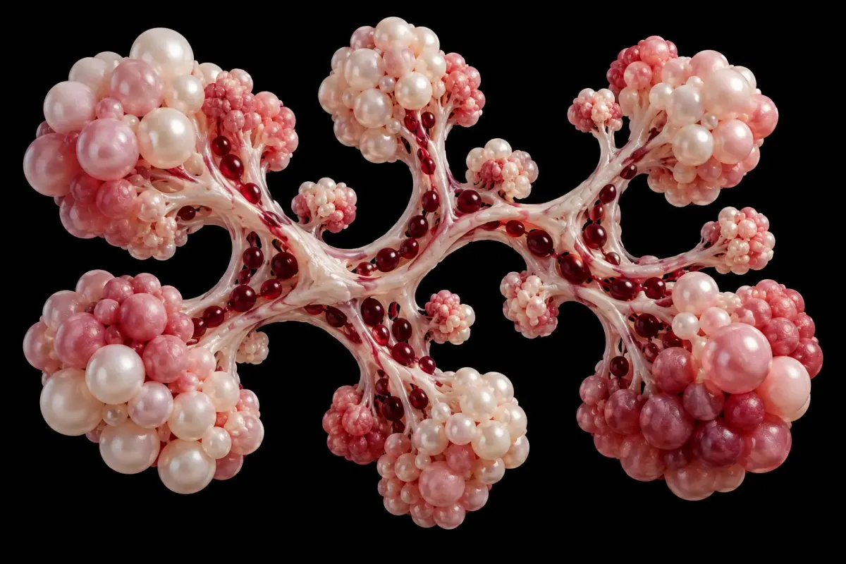
An organoid is a laboratory-grown, three-dimensional arrangement of cells that reproduces selected features of a tissue. It is not necessarily a miniature, fully functioning organ. Here, the units were roughly 200 micrometers across and contained two complementary cell populations: endothelial cells, which line blood vessels, and mural cells, which support their walls.
The researchers began with human pluripotent stem cells—cells capable of developing into many tissue types—and guided their development through a two-stage process. After an initial flat-culture phase, a rotating bioreactor helped the cells aggregate and develop into suspended vascular units. The main protocol took six days. The goal was reproducible organization, not simply a larger number of cells.
The units were not finished arteries ready to plug in. After transplantation, neighboring modules assembled into larger structures. Spaces between them became interconnected channels that could carry blood. This is the distinctive construction idea: organize the cells before delivery, then let the spaces between their modules become part of the circulation.
A blood vessel needs more than cells that look right. It needs an open channel, a stable lining and a connection to circulating blood. These researchers transplanted small, pre-organized groups of vessel-forming cells. In mice, the groups helped create working connections more effectively than loose cells. Whether those connections can remain useful for long periods is still unresolved.
The full paper describes comparisons with corresponding single-cell preparations. This matters: an attractive image of a new vessel would not, by itself, show that pre-assembly improved on a simpler injection. The team also used fluorescent tracers to distinguish vessel-like structures from channels actually reached by the circulation.
Repair involved the host, not just the graft
In the hindlimb experiment, researchers surgically interrupted arterial supply and injected the organoids into the affected muscle. After two weeks, measured perfusion—blood flowing through tissue—reached approximately 40% of the opposite, healthy limb’s level. That was better than the single-cell comparison, but it was not restoration to normal circulation.
The transplanted structures were not acting alone. The researchers observed early recruitment of macrophages, immune cells that can participate in tissue repair. Analyses implicated signaling involving vascular endothelial growth factor A, a protein that encourages blood-vessel growth. Reducing monocyte and macrophage populations with a depletion treatment weakened the organoids’ vascular and perfusion benefits.
That intervention supports a contribution from the host’s repair response. It does not establish that one signaling molecule explains the whole effect. The distinction matters for translation: the same engineered cells may behave differently in a different immune environment.
A working connection still has to last
The clearest durability test came from organoids placed beneath the kidney capsule, a setting used to study graft integration separately from ischemic injury. Blood-carrying networks remained visible at one month. At two months, vascular structures could still be seen, but tracer perfusion was largely absent. By three months, the labeled endothelial networks were no longer detectable. The supplementary follow-up is essential to understanding the result.
That separate graft experiment does not prove that every benefit in injured tissue disappears on the same schedule. Equally, the two-week disease experiments cannot establish lasting recovery. Temporary assistance to the body’s own repair process and permanent replacement of blood vessels are different therapeutic goals.
Most disease experiments used young male immunodeficient mice. Additional mice carrying human immune cells also showed improved perfusion, but graft signals declined over time. Such animals are not substitutes for people with chronic vascular disease. The authors report randomized animal groups and blinded analyses, but no statistical calculation to determine sample sizes in advance. They also disclose patent applications covering the technology.
The advance is a testable design principle: deliver an organized vascular partnership rather than asking isolated cells to assemble one under stress. Its next test is not another beautiful vessel image. It is sustained blood flow, meaningful tissue function and acceptable safety over longer periods in models that better reflect the people a treatment would eventually need to help.
Keep exploring
- A brain implant decodes words and gestures at once — another route from biological signals to useful function.
- Tumor shrinkage and the harder question of more time — why the endpoint matters.
- What a mouse lifespan result can actually establish — reading animal evidence without turning it into a human promise.
AI-assisted. Sources checked.