Some old cells appear to carry a chemical signature that light can read. In aged mouse lung and skin, researchers found a narrow Raman-spectroscopy signal associated with lipid-rich cells expressing p21, a widely used marker of cellular senescence. Their system combined that optical signal with gene activity and spatial location to identify senescent cells without first attaching a fluorescent label.
The peer-reviewed Nature Aging technical report published September 21 is a measurement study, not an anti-aging treatment. It used naturally aged mice, cultured cells and a mouse wound-healing model. The work shows that a cell’s chemistry can add information that gene-expression measurements miss. It does not establish a diagnostic test for people or prove that every cell with the signature is harmful.
Genes describe instructions; Raman light samples chemistry
Cellular senescence is a stress response in which a cell stops dividing but remains biologically active. Senescent cells can help suppress tumors and organize wound repair; when they persist, their secretions can also sustain inflammation and disrupt tissue. That diversity is why no single marker perfectly defines the state.
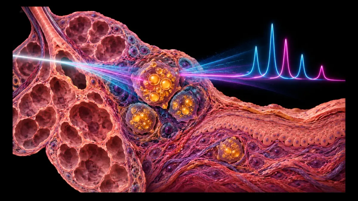
Most cellular maps begin with RNA, the molecules that reflect which genes are being used. Raman spectroscopy asks a different question. A laser interacts with molecular bonds, and a tiny fraction of the scattered light shifts in energy. Those shifts form a spectrum containing information about lipids, proteins, nucleic acids and extracellular material. The method can examine tissue without adding a chemical stain, although interpreting the overlapping signals is difficult.
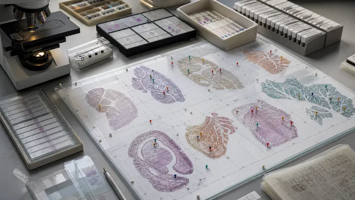
RamanOmics links four layers: single-nucleus RNA sequencing, spatial transcriptomics, hyperspectral Raman imaging and a machine-learning model. The researchers analyzed adjacent slices from lungs and skin of two-month-old and 26-month-old mice. For each tissue and age, they used three animals. The gene-expression dataset included 35,474 lung cells and 12,128 skin cells; the spatial assay targeted a curated panel of 890 genes.
RNA sequencing can tell researchers which biological programs a cell is running, but it usually destroys the cell and does not directly measure its fats or protein shapes. Raman imaging shines light on intact tissue and reads a chemical spectrum. RamanOmics aligns the two views so a spectral pattern can be connected to a cellular state.
Senescence did not look identical across tissues
Old lung and old skin showed different aging programs. In the lung, endothelial and immune cells displayed prominent changes involving inflammation, antigen presentation and extracellular-matrix remodeling. In skin, fibroblasts and epidermal cells changed more strongly, with signals involving metabolism, ion handling and differentiation.
The team then focused on cells positive for p21. Across lung and skin, these cells shared an enhanced Raman band around 1,131–1,135 inverse centimeters, which the authors link to lipid chemistry. That conserved optical feature sat alongside tissue-specific gene programs: lung senescence emphasized matrix remodeling and transforming growth factor-beta signaling, while skin senescence emphasized epidermal differentiation genes including Krt10, Lor and Sbsn.
A machine-learning classifier fused selected Raman and transcriptional features into what the paper calls a multimodal barcode. The point is not that one bright spectral peak replaces biology. It is that chemical and genetic evidence can reinforce each other. A label-free optical readout could eventually help researchers locate candidate senescent cells before choosing which ones to isolate or perturb.
A wound shows why old cells are not one fixed category
In a mouse skin-wound model, senescent cells reactivated epidermal-differentiation programs while the lipid-associated Raman signal increased. That result fits a more complicated view of senescence: the same broad state can participate in repair at one time and become damaging when it accumulates or persists.
The study therefore weakens the idea that senescent cells are a single molecular target waiting for one universal marker. Lung and skin shared part of the chemical signature but differed in the programs surrounding it. A treatment designed simply to remove every p21-positive cell could erase useful states along with harmful ones. The paper does not test such a treatment; it makes the measurement problem more visible.
Its limits matter. The main tissue comparison used three young and three old mice per organ, with two ages standing in for an entire aging trajectory. The spatial transcriptomics panel was selected in advance rather than measuring every transcript. The classifier was developed within the same experimental framework whose signals it combined, so independent laboratories, other organs and blinded prospective samples must show how well it generalizes. Human tissue and live-animal imaging remain future tests.
The most important next result would be functional. Researchers need to show that a Raman-defined state predicts what a cell will do—repair, inflame, recover or resist treatment—better than existing markers alone. If it can, light would become more than a gentler label. It would offer a way to watch the chemistry of aging cells change before destroying the sample to learn what was there.
Keep exploring
- The Human Senescence Atlas turns aging into a mapping problem — why cell identity and location matter before intervention.
- Your organs do not age on one clock — how tissue structure changes on different schedules.
- Semaglutide extended lifespan in mice. What did it prove? — a guide to separating a mouse result from human benefit.
AI-assisted. Sources checked.