The middle-aged male mice had T cells inside their tumors. What they lacked was an equally effective response from those cells. In experiments reported in Nature Aging on October 9, researchers traced part of that gap upstream, to the lymph nodes where matching immune cells can be activated against a tumor.
The finding offers a different way to think about immune aging. A defense can weaken before its cells reach the target, because too few of the right cells are available at the place where recognition begins. The intervention evidence comes from mice; supporting human observations do not establish a treatment for people.
A meeting has to happen before an attack
CD8 T cells can develop into cells that kill infected or cancerous cells. But they are not interchangeable. Different T-cell clones carry different receptors, and only some can recognize a particular fragment of a tumor.
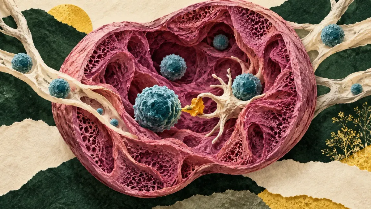
Before activation, these cells are called naive. That does not mean defective. It means they have not yet been activated by the antigen their receptors recognize. In a lymph node receiving material from a tumor, an antigen-presenting cell can display such a fragment to a matching T cell. That encounter can start the response.
Having a diverse repertoire somewhere in the body is therefore not the whole problem. Enough suitable cells must be available locally for an encounter to occur. A smaller pool gives rare, tumor-matching cells fewer opportunities to meet their signal.
In the study's melanoma experiments, middle-aged male and female mice had comparable overall CD8 T-cell infiltration into tumors. Yet the males had fewer cells displaying signs of tumor-specific activation and weaker tumor control. Counting the occupants missed an important difference in what those occupants could do.
The supply shrinks from two directions
The researchers identified two processes reducing the naive CD8 pool. The thymus, the organ that produces new T cells, became less productive with age. At the same time, some naive cells shifted into a state called virtual memory without an encounter with their matching antigen.
That second process matters because a cell can leave the naive pool without simply dying. A total T-cell count can hide a change in the kinds of cells available for a new response. In the mice, earlier thymic decline and faster virtual-memory differentiation combined to produce a more pronounced deficit in males by middle age, alongside smaller lymph nodes.
This is a different question from the CD4-cell remodeling observed in extremely old people. There is no single immune-aging meter: cell type, state, location and the response being tested all matter.
Replenishing the pool helped mice, with an age limit
To test whether the supply bottleneck could be changed, the team used interventions that reduced androgen signaling in male mice. One involved degarelix, a drug that suppresses production of sex hormones. The researchers observed renewed thymic activity and more naive CD8 T cells in lymph nodes.
In middle-aged male mice, this was accompanied by better recognition and control of transplanted melanoma. A direct drug-versus-control tumor-growth comparison involved eight male mice per group. Pretreatment also improved the response to subsequent anti-PD-1 immunotherapy, which releases a brake on immune activity.
The study defined middle-aged mice as 9 to 12 months old and aged mice as older than 18 months. In the older animals, replenishment was weaker and the functional benefit was limited. Raising a cell count did not amount to resetting an old immune system.
The experiments used small groups, and investigators were not blinded to group allocation during experiments or outcome assessment. These were controlled mouse tumor models, including melanoma engineered to carry a model antigen that makes particular immune responses easier to track. They do not reproduce the full variety of cancers in people. Neither the mouse findings nor the use of an existing drug establishes that hormone suppression prevents cancer or reverses immune aging in humans.
Human observations support the question, not the therapy
The human part of the work used existing imaging and immune-cell datasets. It found age- and sex-associated patterns consistent with the mouse mechanism, including differences in lymph-node volume and naive-cell populations. The researchers did not test the intervention in people.
The authors explicitly describe the lymph-node imaging analysis as exploratory. Clinical and anatomical differences can affect the comparisons, and fat within a lymph node complicates the relationship between its apparent size and its immune-cell population. A scan is not a direct count of tumor-recognizing cells.
The result fits a broader lesson from efforts to map aging cells in their tissue context: where cells are, and which state they occupy, can matter as much as whether they are present.
For cancer immunity, the useful distinction is between having defenders and starting a defense. These experiments place one bottleneck at the meeting point: enough still-uncommitted T cells, with the right receptors, must encounter the tumor's signal before an effective response can begin.