
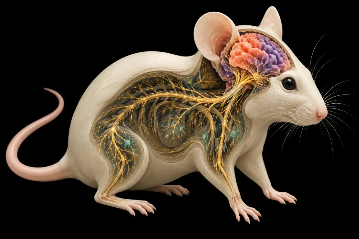
These mice did not receive human brains. They received human cortical organoids—small, stem-cell-derived pieces of developing neural tissue—after researchers genetically removed most of the mouse neurons that would normally form the cortex and hippocampus. The extra space let the grafts grow unusually large, connect with the remaining mouse nervous system and reveal an injury response that ordinary mice did not show.
That is the central result of a peer-reviewed Nature paper published September 16. It is a new experimental platform, not a therapy. The grafts remained developmentally immature and lacked several defining features of an organized mature cortex. Their relationship to the animals’ behavior is not yet established.
The experiment began by making room
Brain organoids are three-dimensional tissues grown from induced pluripotent stem cells—adult cells reprogrammed into a state from which they can develop into many cell types. In a dish, cortical organoids reproduce parts of early human brain development. When transplanted into a rodent brain, they gain blood supply and sensory input, but an intact host cortex limits how far they can expand and which connections they can make.
The Stanford-led team changed the host. It deleted the cohesion gene Esco2 in early cells marked by Emx1, causing most of the developing mouse neocortex and hippocampus to be lost. The animals were also immunodeficient so they would not reject human cells. The researchers call these hosts “apallial” mice and the engrafted animals “xenocortical” mice.
Earlier organoid transplants were like adding a room to an occupied house: the host brain already filled the space and controlled the wiring. Here, researchers removed much of the mouse’s future cortex before birth, then implanted human cortical tissue after birth. The graft had room to expand, but it still developed inside a mouse body and never became a complete human cortex.
Across 29 transplanted mice made with three stem-cell lines, 86.2% showed graft survival by magnetic-resonance imaging two months later. In 14 mice followed between months two and three, graft volume grew about 4.7-fold. In a seven-mouse subset measured at three months, human-derived tissue made up 91.9% of the combined cortical tissue volume.
Those percentages sound dramatic because the mice had first been engineered to lack most native cortical tissue. They do not mean that 91.9% of the whole brain was human, or that the animals possessed a human brain. Subcortical mouse structures and the rest of the mouse nervous system remained.
Human cells connected without becoming a mature cortex
The grafts produced several cortical cell types and extended axons through the host brain and into the spinal cord. Calcium imaging and electrical recordings detected organized activity resembling early developing circuits. Transcriptomic comparisons placed the excitatory neurons at roughly a late-second-trimester human developmental state after 24 weeks of differentiation.
The model also generated a population resembling layer-5 extratelencephalic projection neurons, including cells with features associated with von Economo neurons. These specialized neurons are difficult to produce in organoids kept only in dishes. The paper reports that the relevant gene program was far more common in xenocortical grafts than in a large atlas of in-vitro organoids.
But growth is not organization. The grafts did not form a continuous six-layer cortical plate. They lacked complete regional patterning, a mature transcriptional profile and the normal representation of inhibitory neurons. Human neurons mature much more slowly than mouse neurons; that developmental mismatch may put a ceiling on integration.
An injury test shows both promise and ambiguity
The researchers exposed control, apallial and xenocortical mice to low oxygen. Human graft cells showed substantial injury, while ordinary mouse brains were more resilient under the tested conditions. Afterward, the xenocortical mice changed how they supported their weight while walking. That gives researchers a path from a human-cell injury to an observable animal-level readout.
It does not prove that the human graft caused a particular behavior. The authors explicitly say it remains unclear whether graft-derived neurons are necessary or sufficient for the observed differences. Many behavioral groups were small, and the animals began with an extraordinary developmental alteration. Their preserved basic movement shows that subcortical systems can carry more function than a cortex-centered picture might suggest; it does not make these mice ordinary.
The ethical question therefore grows with the scientific capability. More mature, better organized grafts may model disease more faithfully, but they could also create properties that deserve new scrutiny. The authors call for early guidance before work pursues later maturation, cortical folding, balanced excitatory and inhibitory networks, embryonic transplantation or non-human primate hosts.
There is also a disclosed commercial interest: Stanford holds patents on cortical-organoid generation and a provisional patent application for organoid transplantation, listing several study authors as inventors.
The next decisive experiments are pathway-specific. Researchers need to switch defined graft circuits on or off and show whether a particular animal readout follows. They also need to test disease-associated human cells across multiple donor lines. The platform’s value is not that it blurred the boundary between species. It is that it created enough living human cortical tissue to make previously inaccessible questions measurable—while making the limits impossible to ignore.
Keep exploring
- A brain implant decodes words and gestures at once — another case where neural signals need careful interpretation.
- These cell clusters give blood a new route — how pre-organized tissue changes an animal experiment.
- This fly wiring map doesn’t stop at the neck — what a complete circuit map can and cannot reveal.
AI-assisted. Sources checked.